Thebault Farriery
X-Ray Block top
X-Ray Block top
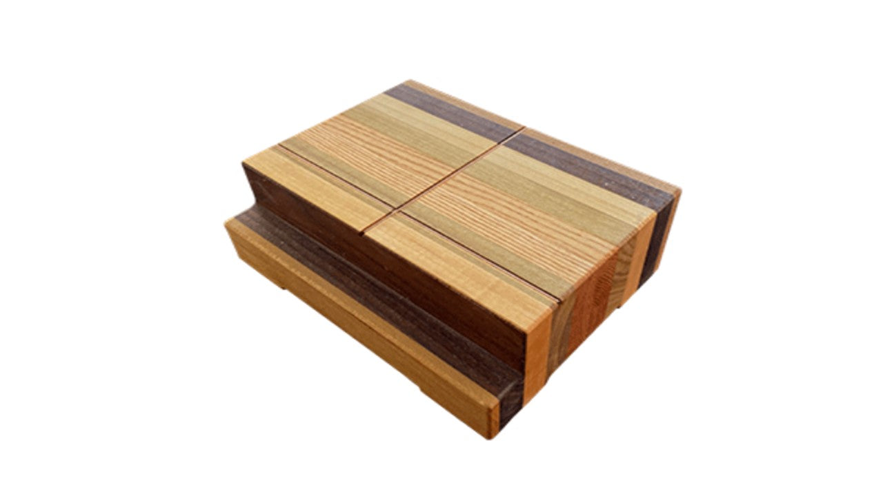
These wooden blocks are designed by Dr. Redden to give you a consistent way to achieve precise finger positioning for lateral and DP views. Dr. Redden strives to get all possible information from his film and he has developed a very methodical and disciplined protocol designed for every x-ray examination. The wooden positioning block has multiple uses and provides many valuable diagnostic positions.
Dr. Redden updated the blocks by staggering the cross wires so the intersection was closer to the cassette. The higher blocks (NR-1) have a cassette shelf.
Unique Block Features:
- Sold individually or in pairs depending on demand. Dr. Redden finds that using two blocks ensures an accurate depiction of radiographic balance (extremely valuable views for farriers).
- Two blocks simplify the task of x-raying extremely lame horses. The sound foot can be quickly placed on a block and the other foot follows.
- The height of the blocks is specific to each machine. The height of the main beam should be approximately 3/4 inch (19.05 mm) above the block. This ensures true lateral measurement of sole depth, palmar angle, medial/lateral balance and distal horn/lamellar area. Exception: Saddlebreds, Walking Horses or others with an exceptionally long hoof pack or capsule.
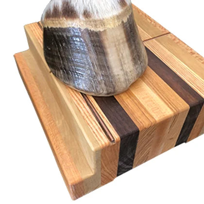
- A cross wire embedded in the face of each block clearly delineates the ground surface in the side projection and also in the DP view; very useful load zone marker for bare foot. Dr. Redden uses Intropaste to clearly define the horn wall.
- Dr. Redden advises placing the foot so that it rests on the edge of the block touching the cassette. The alignment of the foot should be such that the line formed by the center of the heel and the center of the toe is parallel to the long vertical crosshairs. Ideally, the short horizontal reticle runs through the center of the foot and gives you a direct line to follow for your x-ray beam. Note: If the horse goes in or out, adjust your block accordingly to get the best shot.
Consult Dr. Redden's monograph, Radiography of the Equine Foot (Series One) to learn more about the radiographic technique.
NR-1 measures
- Height: 3 3/8 inches (85.7 mm)
- Width: 7 1/16 inches (179.4mm)
- Length: 9 inches (228.6mm)
- Compatible with MinXray HF100+, HF100/30, HF100/40 or any X-ray device with a primary beam height of 4 to 4 1/4 inches.
NR-2 measures
- Height: 2 5/16 inches (58.7 mm)
- Width: 7 1/16 inches (179.4mm)
- Length: 9 inches (228.6mm)
- Compatible with MinXray TR8020, TR80, TR90, HF8015, HF8015+ or any X-ray device with a primary beam height of 4 to 4 1/4 inches.


